 Skip to content
Which image represents cytokinesis in an animal cell – Thus, option d is correct. During male meiosis in humans, for example, all 4 cells at the end of meiosis have the same size, and relative number of organelles. Web in this review, we provide an overview of four topics in animal cell cytokinesis: Telophase & cytokinesis, 250x, whitefish embryo. Web first, it is important to note that cytokinesis is the process by which a cell divides into two daughter cells, so any image of cytokinesis should clearly show two separate cells emerging from the original cell. Web which image represents cytokinesis in an animal cell c which step of mitosis involves the condesing of dna into chromosomes? 10.1101/cshperspect.a015834 abstract cell division ends with the physical separation of the two daughter cells, a process known as cytokinesis. Meanwhile, cytokinesis in animal cells produces new cell membranes from the endoplasmic reticulum. Web in this review, we provide an overview of four topics in animal cell cytokinesis: Web which image represents cytokinesis in an animal cell get the answers you need, now!
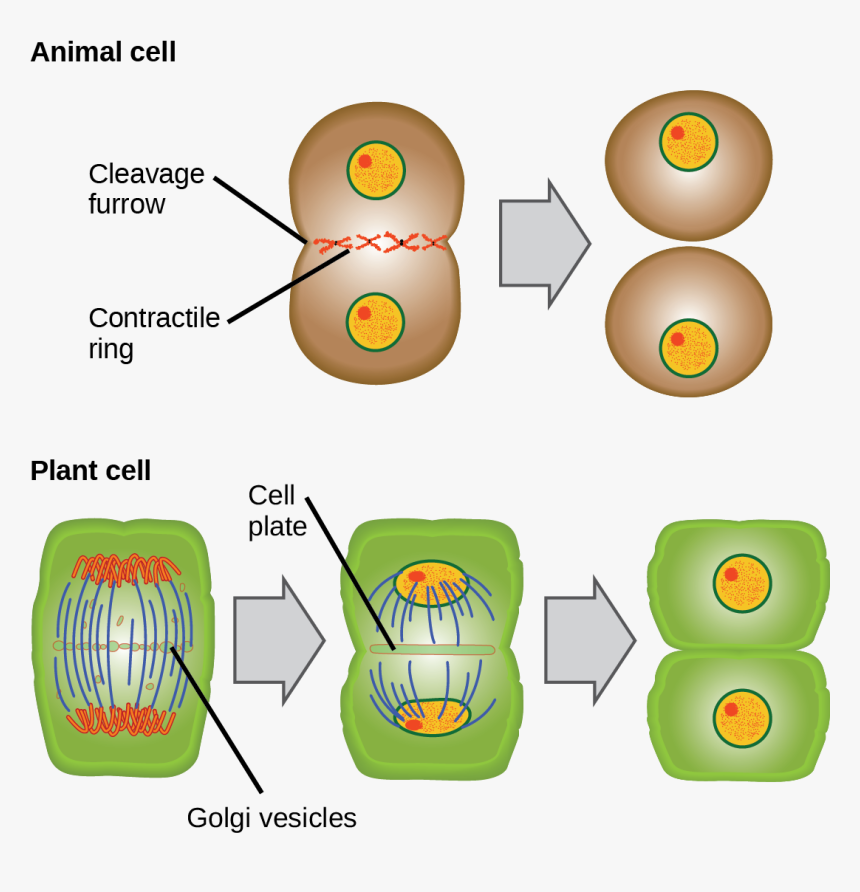
Many of these cells are undergoing division since the root tip grows quickly and requires more cells. (a) signaling between the anaphase spindle and cortex, (b) the mechanics of cortical remodeling, (c) abscission, and (d) regulation of cytokinesis by the cell cycle machinery. Interphase, mitosis, cytokinesis 1 multiple choice option arthur made a mistake labeling the diagram of the three stages of the cell cycle. Importantly, cytokinesis takes place differently in animal and plant cells. Web a plant cell contains a large, singular vacuole that is used for storage and maintaining the shape of the cell.

Cytokinesis In Plant Cells slidesharedocs
Web in this review, we provide an overview of four topics in animal cell cytokinesis: During male meiosis in humans, for example, all 4 cells at the end of meiosis have the same size, and relative number of organelles. 10.1101/cshperspect.a015834 abstract cell division ends with the physical separation of the two daughter cells, a process known as cytokinesis. Cytokinesis, or “cell motion,” is the second main stage of the mitotic phase during which cell division is completed via the physical separation of the cytoplasmic components into two daughter cells. Plant cells can’t be divided like this because they have a cell wall and are too stiff. In particular, the image shows the nucleus of. Particular functions demand various deviations from the process of symmetrical cytokinesis; Cytokinesis 3 multiple choice options which are the main stages of the cell cycle? Web in this review, we provide an overview of four topics in animal cell cytokinesis: 4.which image represents cytokinesis in an animal cell?
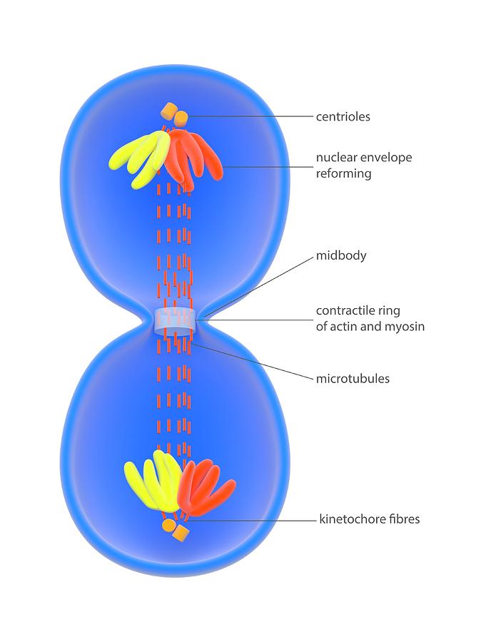
Plant cells have a cell wall, as well as a cell membrane. Web midbody formation and structure in late cytokinesis.

10.2B The Mitotic Phase and the G0 Phase Biology LibreTexts
During male meiosis in humans, for example, all 4 cells at the end of meiosis have the same size, and relative number of organelles. For example in oogenesis in animals the ovum takes almost all the cytoplasm and organelles. Cytokinesis 3 multiple choice options which are the main stages of the cell cycle? Plant cells have a cell wall, as well as a cell membrane. In contrast, animal cells have many, smaller vacuoles. Web which part of the cell cycle does this image represents in an animal cell? Hela cells were fixed and stained to detect tubulin and dna. Web which image represents cytokinesis in an animal cell c which step of mitosis involves the condesing of dna into chromosomes? Chromosomes, cleavage furrow, spindle fibers, and daughter cells. Web suzanne wakim & mandeep grewal butte college table of contents the forms of dna divide and split can you guess what this colorful image represents?
Web in this review, we provide an overview of four topics in animal cell cytokinesis: Cytokinesis usually begins just as mitosis is ending, with a little overlap.

Topics Which Image Represents Cytokinesis In An Animal Cell Latest
Web midbody formation and structure in late cytokinesis. It shows a eukaryotic cell during the process of cell division. Web in animal cells, cytokinesis is contractile, pinching the cell in two like a coin purse with a drawstring. In contrast, animal cells have many, smaller vacuoles. In particular, the image shows the nucleus of. Hi it is b relly it is i am not lyinggggggggg Web study with quizlet and memorize flashcards containing terms like which step of mitosis involves the nucleus splitting and nuclear membranes forming around each new nucleus?, which image represents cytokinesis in an animal cell?, jacqueline is trying to draw an image of a cell in telophase. Web a plant cell contains a large, singular vacuole that is used for storage and maintaining the shape of the cell. Cytokinesis 3 multiple choice options which are the main stages of the cell cycle? When it comes to the spindle, the spindle middle part stays active and vigorous during cytokinesis in plant cells, but in animal cells, the cytokinesis spindle.
A crucial function of cytokinesis is to divide the cell in half and make sure that one nucleus is placed in each daughter cell. Web in this review, we provide an overview of four topics in animal cell cytokinesis:
Cytokinesis In Animal Cells, HD Png Download kindpng
( a) signaling between the anaphase spindle and cortex, ( b) the mechanics of cortical remodeling, ( c) abscission, and ( d) regulation of cytokinesis by the cell cycle machinery. Telophase & cytokinesis, 250x, whitefish embryo. 10.1101/cshperspect.a015834 abstract cell division ends with the physical separation of the two daughter cells, a process known as cytokinesis. Hi it is b relly it is i am not lyinggggggggg Plant cells have a cell wall, as well as a cell membrane. Web in cytokinesis, the cytoplasm of the cell is split in two, making two new cells. Web first, it is important to note that cytokinesis is the process by which a cell divides into two daughter cells, so any image of cytokinesis should clearly show two separate cells emerging from the original cell. Web suzanne wakim & mandeep grewal butte college table of contents the forms of dna divide and split can you guess what this colorful image represents? Meanwhile, cytokinesis in animal cells produces new cell membranes from the endoplasmic reticulum. Hela cells were fixed and stained to detect tubulin and dna.
Plant cells can’t be divided like this because they have a cell wall and are too stiff. Interphase, mitosis, cytokinesis 1 multiple choice option arthur made a mistake labeling the diagram of the three stages of the cell cycle.

Cytokinesis The process that follows the last stage of mitosis. With
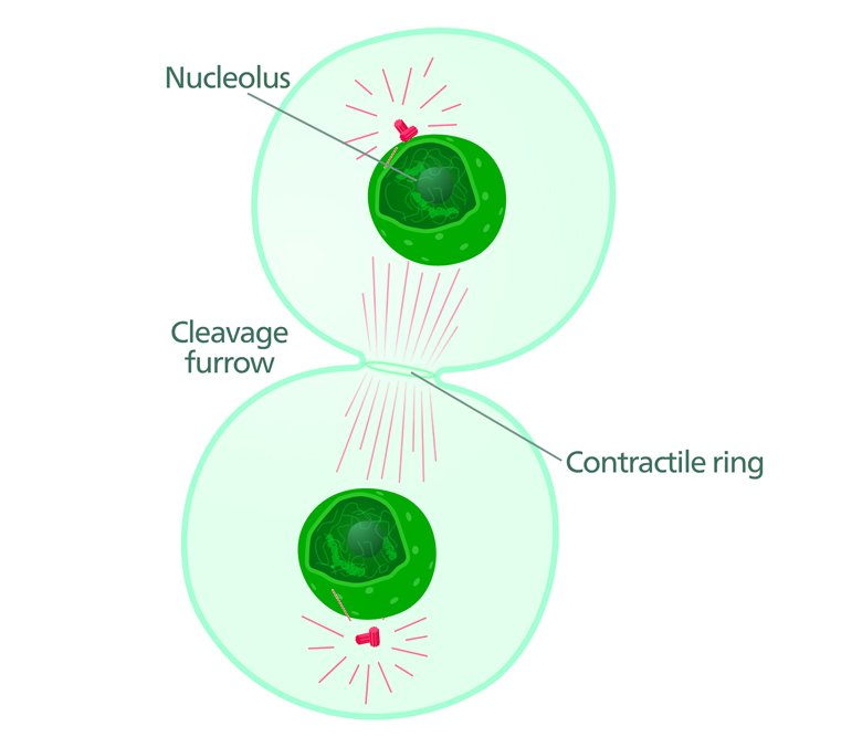
Which describes what she should draw? Which image represents cytokinesis in a plant cell? Prophase a student is looking through microscope at some cells of an onion root tip. ( a) signaling between the anaphase spindle and cortex, ( b) the mechanics of cortical remodeling, ( c) abscission, and ( d) regulation of cytokinesis by the cell cycle machinery. Web identify the characteristics of cytokinesis. Hi it is b relly it is i am not lyinggggggggg Cytokinesis 3 multiple choice options which are the main stages of the cell cycle? Meanwhile, cytokinesis in animal cells produces new cell membranes from the endoplasmic reticulum. Web in animal cells, cytokinesis is contractile, pinching the cell in two like a coin purse with a drawstring. Web which image represents cytokinesis in an animal cell get the answers you need, now!
Plant cells have a cell wall, as well as a cell membrane. Where is the error in the diagram?
What Forms In Animal Cells During Cytokinesis Cytokinesis in animal
Interphase, mitosis, cytokinesis 1 multiple choice option arthur made a mistake labeling the diagram of the three stages of the cell cycle. When it comes to the spindle, the spindle middle part stays active and vigorous during cytokinesis in plant cells, but in animal cells, the cytokinesis spindle. A crucial function of cytokinesis is to divide the cell in half and make sure that one nucleus is placed in each daughter cell. Cytokinesis 3 multiple choice options which are the main stages of the cell cycle? Telophase & cytokinesis, 250x, whitefish embryo. Web after the completion of the telophase and cytokinesis, each daughter cell enters the interphase of the cell cycle. Cytokinesis usually begins just as mitosis is ending, with a little overlap. Which image represents cytokinesis in a plant cell? Importantly, cytokinesis takes place differently in animal and plant cells. This gives the plant cell its unique rectangular shape.
Hi it is b relly it is i am not lyinggggggggg Web study with quizlet and memorize flashcards containing terms like which step of mitosis involves the nucleus splitting and nuclear membranes forming around each new nucleus?, which image represents cytokinesis in an animal cell?, jacqueline is trying to draw an image of a cell in telophase.
Cytokinesis In Animal Cells Photograph by Science Photo Library Fine
During male meiosis in humans, for example, all 4 cells at the end of meiosis have the same size, and relative number of organelles. It shows a eukaryotic cell during the process of cell division. Hi it is b relly it is i am not lyinggggggggg Web suzanne wakim & mandeep grewal butte college table of contents the forms of dna divide and split can you guess what this colorful image represents? Plant cells can’t be divided like this because they have a cell wall and are too stiff. Which describes what she should draw? Web which part of the cell cycle does this image represents in an animal cell? (a) signaling between the anaphase spindle and cortex, (b) the mechanics of cortical remodeling, (c) abscission, and (d) regulation of cytokinesis by the cell cycle machinery. Web in animal cells, cytokinesis is contractile, pinching the cell in two like a coin purse with a drawstring. In plants, the cell wall surrounds the cell membrane.
Web a plant cell contains a large, singular vacuole that is used for storage and maintaining the shape of the cell. This final event ensures that nuclear and cytoplasmic contents are accurately partitioned between the two nascent cells.
Meanwhile, cytokinesis in animal cells produces new cell membranes from the endoplasmic reticulum. Web midbody formation and structure in late cytokinesis. Hela cells were fixed and stained to detect tubulin and dna. Web cells can divide evenly, known as symmetrical cytokinesis, or one of the cells can retain a majority of the cytoplasm. The “drawstring” is a band of filaments made of a protein called actin, and the pinch crease is known as the cleavage furrow. Web which part of the cell cycle does this image represents in an animal cell? Cytokinesis, or “cell motion,” is the second main stage of the mitotic phase during which cell division is completed via the physical separation of the cytoplasmic components into two daughter cells.
Telophase & cytokinesis, 250x, whitefish embryo. 4.which image represents cytokinesis in an animal cell? Thus, option d is correct. Plant cells have a cell wall, as well as a cell membrane. Hi it is b relly it is i am not lyinggggggggg 10.1101/cshperspect.a015834 abstract cell division ends with the physical separation of the two daughter cells, a process known as cytokinesis.

“Printable Calendar is a website that provides high-quality and customizable calendars for individuals and businesses. Founded in 2022, the website offers many printable calendars to help people stay organized and manage their time effectively.
Our team of experienced professionals is passionate about creating calendars that are not only functional but also visually appealing. We understand the importance of time management in today’s fast-paced world and strive to make it easier for our customers to plan and schedule their daily activities.
At Printable Calendar, we believe in offering our customers the best possible experience. We constantly update our website with new designs and features to ensure our customers can access the latest and most innovative calendars. We also provide excellent customer support to ensure our customers can get their help whenever needed.
Whether you’re looking for a monthly, weekly, or yearly calendar, Printable Calendar covers you. Our calendars are available in various formats and sizes, making choosing the one that best suits your needs easy. So why wait? Visit Printable Calendar today and start organizing your life!”


![BASD Calendar: Best Online Calendar for [Target Audience/Use Case]](https://lh3.googleusercontent.com/xsJ3CDJGVjb1sE6NU2BFvJuoFDpvPi0m7YLFiMvCLkLY0nTagixlDlFEQZAf7JF8Ijc=h900 "BASD Calendar: Best Online Calendar for [Target Audience/Use Case]")