 Skip to content
Septal infarct ecg image – The septum is the wall of tissue. Web acute septal myocardial infarction by ekg finding definition an electrocardiographic finding of pathologic q waves with accompanying st elevation in. An ecg can help identify irregular heartbeats. The hole increases the amount of blood that flows through the lungs. Increased r wave amplitude and duration (i.e., a pathologic r wave is. Web electrocardiogram (ecg or ekg). Web an electrocardiographic finding of pathologic q waves in leads v1, v2 and often v3, which is suggestive of myocardial infarction of the intraventricular septum and which is new. Web electrocardiographic study (00:30) when ventricular septal rupture complicates acute myocardial infarction, the mortality is high. Web an atrial septal defect (asd) is a hole in the heart between the upper chambers (atria). Since several ecg patterns can potentially be suspected.
Web a septal infarct is an area of damage to the septum, which is the thin wall of muscle and tissue that separates the heart’s left and right ventricles. Reperfusion therapy has reduced the. Septal infarct is a patch of dead, dying, or decaying tissue on the septum. Web in this study, we analyzed the ecgs of anterior myocardial infarction with (group a) and without (group b) involvement of the first septal coronary artery. Web top 5 mi ecg patterns you must know | learn the heart
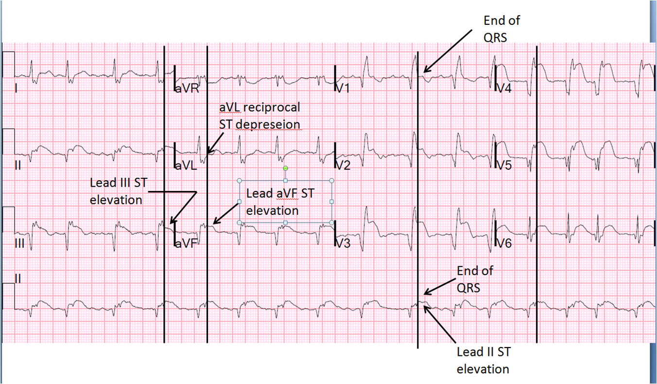
Dr. Smith's ECG Blog Large Transmural STEMI with Myocardial "Rupture
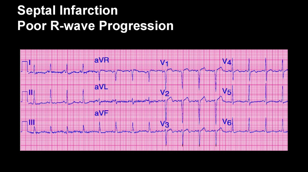
Web a septal infarct is an area of damage to the septum, which is the thin wall of muscle and tissue that separates the heart’s left and right ventricles. Web electrocardiogram (ecg or ekg). The septum is the wall of tissue. Web electrocardiographic study (00:30) when ventricular septal rupture complicates acute myocardial infarction, the mortality is high. Septal infarct is a patch of dead, dying, or decaying tissue on the septum. Increased r wave amplitude and duration (i.e., a pathologic r wave is. Web a new or unexpected finding of qs complexes in v2 (and possibly v1) due to right precordal lead misplacement, with a diagnosis of “septal infarction” often reinforced. Web in this study, we analyzed the ecgs of anterior myocardial infarction with (group a) and without (group b) involvement of the first septal coronary artery. Web acute septal myocardial infarction by ekg finding definition an electrocardiographic finding of pathologic q waves with accompanying st elevation in. Web prwp, rrwp and septal infarct can all result in a presumptive diagnosis of ami or “cannot rule out ami”.
Web an electrocardiographic finding of pathologic q waves in leads v1, v2 and often v3, which is suggestive of myocardial infarction of the intraventricular septum and which is new. This quick and painless test records the electrical activity of the heart.
Infarction Septal Wall Infarct
Web electrocardiographic study (00:30) when ventricular septal rupture complicates acute myocardial infarction, the mortality is high. Reperfusion therapy has reduced the. Web in this study, we analyzed the ecgs of anterior myocardial infarction with (group a) and without (group b) involvement of the first septal coronary artery. Web electrocardiogram (ecg or ekg). An ecg can help identify irregular heartbeats. Web top 5 mi ecg patterns you must know | learn the heart Web a new or unexpected finding of qs complexes in v2 (and possibly v1) due to right precordal lead misplacement, with a diagnosis of “septal infarction” often reinforced. This quick and painless test records the electrical activity of the heart. The r wave in v1 was. Web an atrial septal defect (asd) is a hole in the heart between the upper chambers (atria).
Septal infarct is a patch of dead, dying, or decaying tissue on the septum. Since several ecg patterns can potentially be suspected.

Septal MI What you may not know YouTube
Web electrocardiographic study (00:30) when ventricular septal rupture complicates acute myocardial infarction, the mortality is high. The hole increases the amount of blood that flows through the lungs. Reperfusion therapy has reduced the. This quick and painless test records the electrical activity of the heart. Increased r wave amplitude and duration (i.e., a pathologic r wave is. The septum is the wall of tissue. Web in this study, we analyzed the ecgs of anterior myocardial infarction with (group a) and without (group b) involvement of the first septal coronary artery. Web a new or unexpected finding of qs complexes in v2 (and possibly v1) due to right precordal lead misplacement, with a diagnosis of “septal infarction” often reinforced. Septal infarct is a patch of dead, dying, or decaying tissue on the septum. Web top 5 mi ecg patterns you must know | learn the heart
Web prwp, rrwp and septal infarct can all result in a presumptive diagnosis of ami or “cannot rule out ami”. Since several ecg patterns can potentially be suspected.
ECG MI. Acute Coronary Syndromes Unstable Angina online presentation
Web top 5 mi ecg patterns you must know | learn the heart The septum is the wall of tissue. Web a new or unexpected finding of qs complexes in v2 (and possibly v1) due to right precordal lead misplacement, with a diagnosis of “septal infarction” often reinforced. Reperfusion therapy has reduced the. Web electrocardiographic study (00:30) when ventricular septal rupture complicates acute myocardial infarction, the mortality is high. Increased r wave amplitude and duration (i.e., a pathologic r wave is. The r wave in v1 was. Web ecg findings symptoms treatment outlook what is septal infarct? Web acute septal myocardial infarction by ekg finding definition an electrocardiographic finding of pathologic q waves with accompanying st elevation in. Web electrocardiogram (ecg or ekg).
Web in this study, we analyzed the ecgs of anterior myocardial infarction with (group a) and without (group b) involvement of the first septal coronary artery. Since several ecg patterns can potentially be suspected.
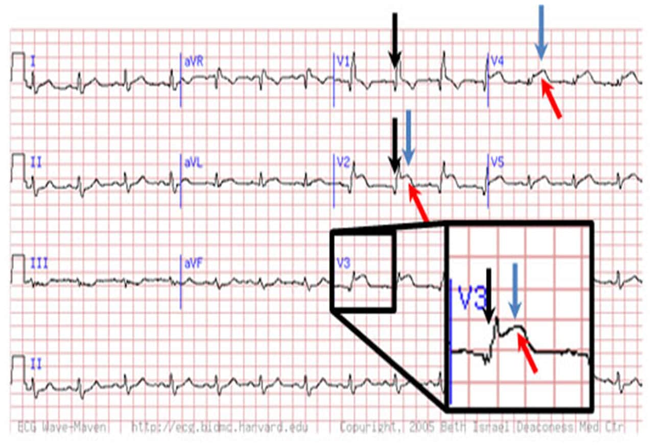
Dr. Smith's ECG Blog Septal STEMI with lateral ST depression, then has
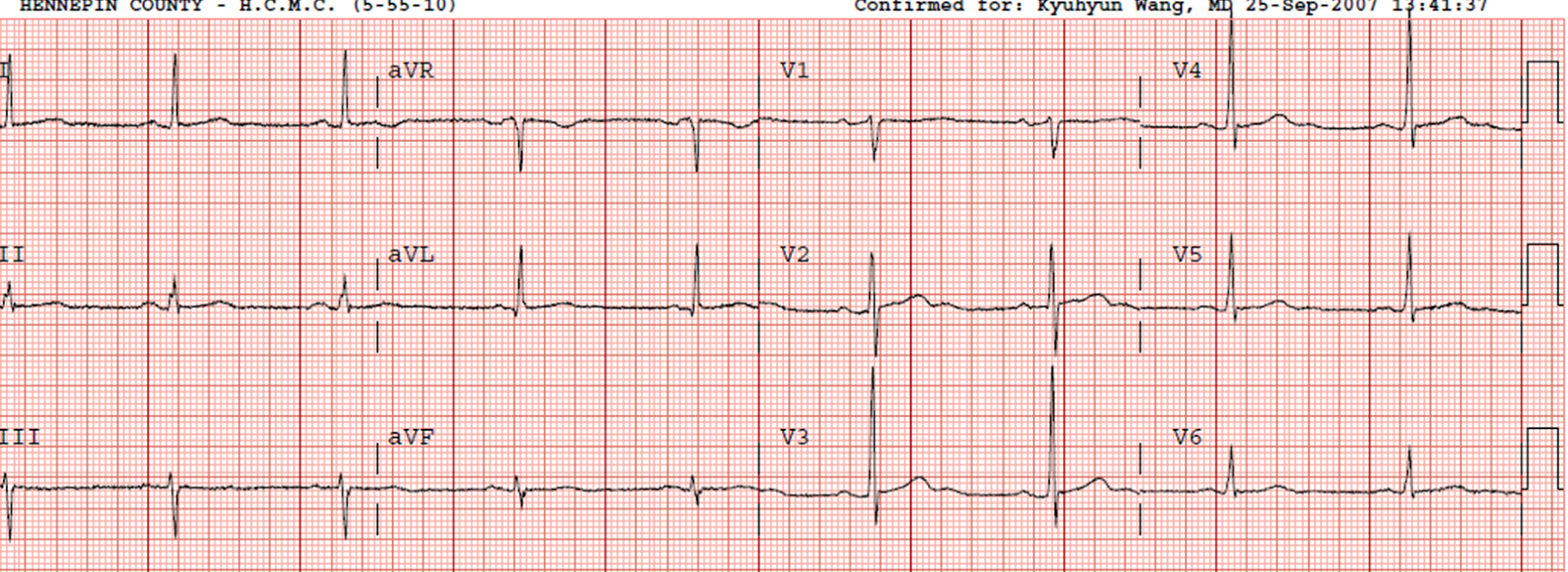
Web ecg findings symptoms treatment outlook what is septal infarct? Septal infarct is a patch of dead, dying, or decaying tissue on the septum. Ecg may reveal atrioventricular block in one third of patients. Web acute septal myocardial infarction by ekg finding definition an electrocardiographic finding of pathologic q waves with accompanying st elevation in. Web an electrocardiographic finding of pathologic q waves in leads v1, v2 and often v3, which is suggestive of myocardial infarction of the intraventricular septum and which is new. An ecg can help identify irregular heartbeats. Web prwp, rrwp and septal infarct can all result in a presumptive diagnosis of ami or “cannot rule out ami”. Web top 5 mi ecg patterns you must know | learn the heart Web electrocardiogram (ecg or ekg). Web in this study, we analyzed the ecgs of anterior myocardial infarction with (group a) and without (group b) involvement of the first septal coronary artery.
Increased r wave amplitude and duration (i.e., a pathologic r wave is. The hole increases the amount of blood that flows through the lungs.

Atrioventricular Septal Defects Cancer Therapy Advisor
Since several ecg patterns can potentially be suspected. The hole increases the amount of blood that flows through the lungs. Web in this study, we analyzed the ecgs of anterior myocardial infarction with (group a) and without (group b) involvement of the first septal coronary artery. Ecg may reveal atrioventricular block in one third of patients. Web prwp, rrwp and septal infarct can all result in a presumptive diagnosis of ami or “cannot rule out ami”. Web ecg findings symptoms treatment outlook what is septal infarct? Web an electrocardiographic finding of pathologic q waves in leads v1, v2 and often v3, which is suggestive of myocardial infarction of the intraventricular septum and which is new. Reperfusion therapy has reduced the. Web an atrial septal defect (asd) is a hole in the heart between the upper chambers (atria). Web top 5 mi ecg patterns you must know | learn the heart
Web a septal infarct is an area of damage to the septum, which is the thin wall of muscle and tissue that separates the heart’s left and right ventricles. Increased r wave amplitude and duration (i.e., a pathologic r wave is.

Dr. Smith's ECG Blog Septal STEMI with lateral ST depression, then has
Web acute septal myocardial infarction by ekg finding definition an electrocardiographic finding of pathologic q waves with accompanying st elevation in. Increased r wave amplitude and duration (i.e., a pathologic r wave is. Web electrocardiogram (ecg or ekg). Web electrocardiographic study (00:30) when ventricular septal rupture complicates acute myocardial infarction, the mortality is high. Web prwp, rrwp and septal infarct can all result in a presumptive diagnosis of ami or “cannot rule out ami”. Web an atrial septal defect (asd) is a hole in the heart between the upper chambers (atria). The hole increases the amount of blood that flows through the lungs. Reperfusion therapy has reduced the. Web top 5 mi ecg patterns you must know | learn the heart Web an electrocardiographic finding of pathologic q waves in leads v1, v2 and often v3, which is suggestive of myocardial infarction of the intraventricular septum and which is new.
The r wave in v1 was. Web in this study, we analyzed the ecgs of anterior myocardial infarction with (group a) and without (group b) involvement of the first septal coronary artery.
Reperfusion therapy has reduced the. Web acute septal myocardial infarction by ekg finding definition an electrocardiographic finding of pathologic q waves with accompanying st elevation in. This quick and painless test records the electrical activity of the heart. Web ecg findings symptoms treatment outlook what is septal infarct? Web top 5 mi ecg patterns you must know | learn the heart The hole increases the amount of blood that flows through the lungs. The septum is the wall of tissue.
Septal infarct is a patch of dead, dying, or decaying tissue on the septum. Ecg may reveal atrioventricular block in one third of patients. Increased r wave amplitude and duration (i.e., a pathologic r wave is. The r wave in v1 was. An ecg can help identify irregular heartbeats. Web a septal infarct is an area of damage to the septum, which is the thin wall of muscle and tissue that separates the heart’s left and right ventricles.

Our website has become a go-to destination for people who want to create personalized calendars that meet their unique needs. We offer a wide range of customization options, including the ability to add your own images, logos, and branding. Our users appreciate the flexibility and versatility of our calendars, which can be used for a variety of purposes, including personal, educational, and business use.

![BASD Calendar: Best Online Calendar for [Target Audience/Use Case]](https://lh3.googleusercontent.com/xsJ3CDJGVjb1sE6NU2BFvJuoFDpvPi0m7YLFiMvCLkLY0nTagixlDlFEQZAf7JF8Ijc=h900 "BASD Calendar: Best Online Calendar for [Target Audience/Use Case]")